Etiology
A pituitary adenoma is a benign, neoplastic proliferation of epithelial cells of the pituitary gland. These cells typically demonstrate both autonomous growth and function. The most common pituitary adenomas in veterinary medicine are microscopic tumors of the basophilic or chromophobic cells of the pars intermedia and the pars distalis. Progressive proliferation of these cells result in secretion of excessive amounts of ACTH, which increases cortisol production by the adrenal gland causing hyperplasia of the gland and signs of pituitary dependent hyperadrenocorticism. Once the dimensions of these tumors exceed 10 mm in diameter they are considered macroadenomas.Pathophysiology
Symptoms from pituitary microadenomas occur secondary to the excessive production of various pituitary hormones. Macroadenomas may also cause symptoms from excessive hormone production but can also result in neurologic symptoms that develop in response to the compression of surrounding central nervous system structures.Clinical Signs
Mental dullness, disorientation, blindness, head pressing, behavioral changes and seizures may occur secondary to direct compression of the overlying diencephalon or the adjacent optic chiasm. Increases in intracranial pressure due to the mass or associated vasogenic edema may also contribute to the general deterioration in mental status.Diagnostic Tests
MRI is the preferred advanced diagnostic imaging modality. On MRI images pituitary macroadenomas are usually isointense on T1 weighted images, hyperintense on T2 weighted images and demonstrate a uniform enhancement following contrast administration. Bleeding within the tumor (i.e., pituitary apoplexy) will lead to regions of markedly reduced GRE T2* signal (see images below). Occasionally pituitary macroadenomas demonstrate internal cystic regions that are uniformly T1 hypointense and T2 hyperintense and fail to enhance following contrast administration (see images below). Pituitary macroadenomas can also be diagnosed using CT of the head and typically appear as a variably contrast enhancing soft tissue dense mass originating within the pituitary fossa (see image below).Therapy
Radiation therapy represents the only definitive therapy option currently available in the United States.Image Gallery
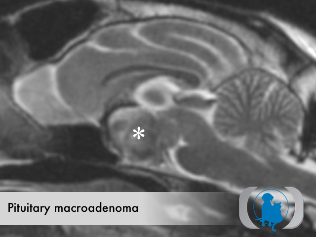
Sagittal T2 weighted magnetic resonance image (MRI) of the brain of a 12 year old neutered male DSH cat with a hemorrhagic pituitary macroadenoma (white asterisk).
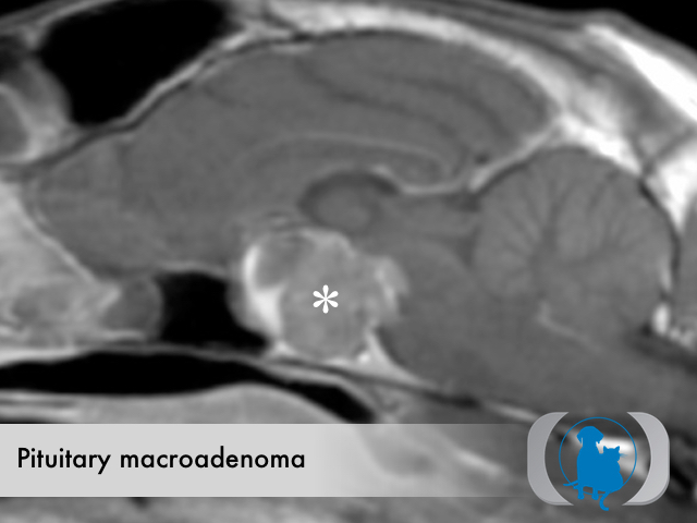
Sagittal T1 weighted post contrast magnetic resonance image (MRI) of the brain of a 12 year old neutered male DSH cat with a hemorrhagic pituitary macroadenoma (white asterisk).
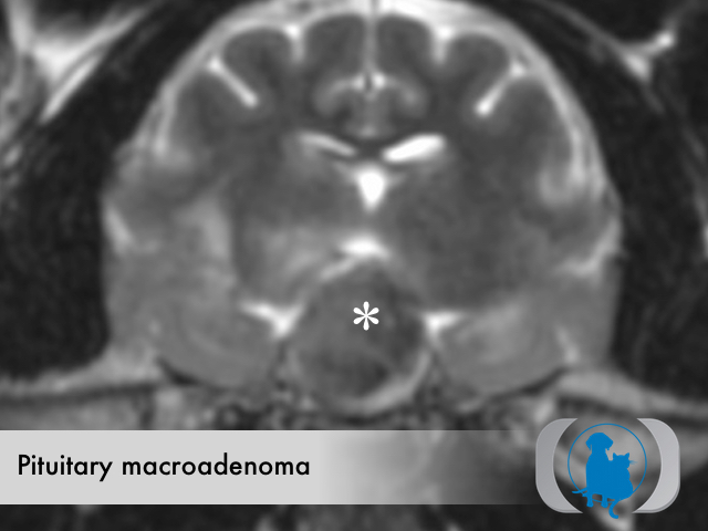
Axial T2 weighted magnetic resonance image (MRI) of the brain of a 12 year old neutered male DSH cat with a hemorrhagic pituitary macroadenoma (white asterisk).
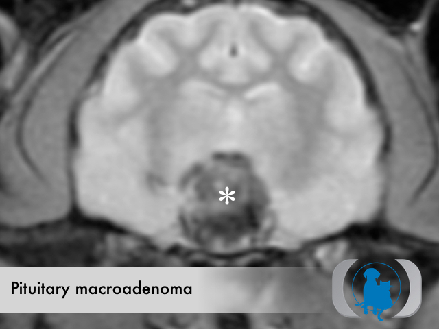
Axial T2* weighted magnetic resonance image (MRI) of the brain of a 12 year old neutered male DSH cat with a hemorrhagic pituitary macroadenoma (white asterisk). Note the extensive regions of markedly decreased signal consistent with hemorrhage on this sequence.
Axial T1 weighted post contrast magnetic resonance image (MRI) of the brain of a 12 year old neutered male DSH cat with a hemorrhagic pituitary macroadenoma (white asterisk).
Dorsal T1 weighted post contrast magnetic resonance image (MRI) of the brain of a 12 year old neutered male DSH cat with a hemorrhagic pituitary macroadenoma (white asterisk).
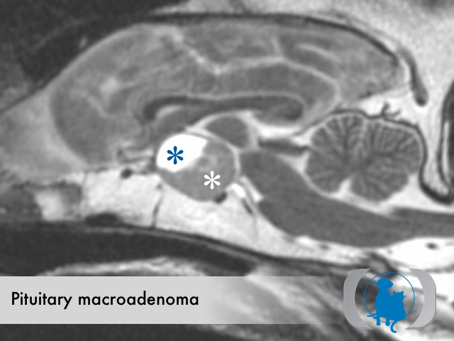
Sagittal T2 weighted magnetic resonance image (MRI) of the brain of a 6 year old neutered male Labrador Retriever dog with a partially cystic (blue asterisk) pituitary macroadenoma (white asterisk).
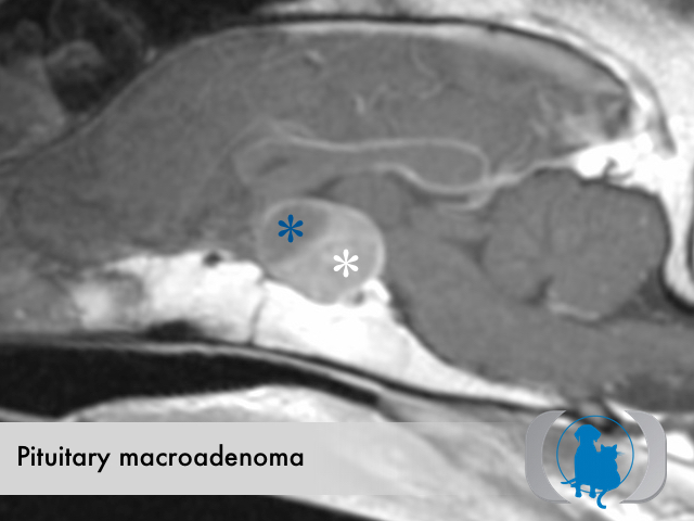
Sagittal T1 weighted post contrast magnetic resonance image (MRI) of the brain of a 6 year old neutered male Labrador Retriever dog with a partially cystic (blue asterisk) pituitary macroadenoma (white asterisk).
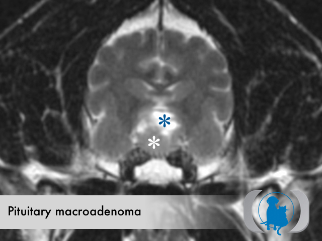
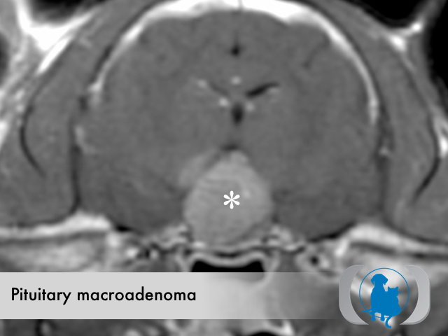
Axial T2 weighted magnetic resonance image (MRI) of the brain of a 6 year old neutered male Labrador Retriever dog with a partially cystic (blue asterisk) pituitary macroadenoma (white asterisk).
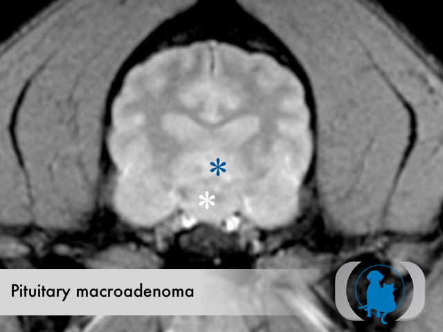
Axial T2* weighted magnetic resonance image (MRI) of the brain of a 6 year old neutered male Labrador Retriever dog with a partially cystic (blue asterisk) pituitary macroadenoma (white asterisk).
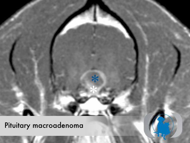
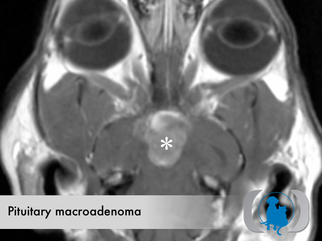
Axial T1 weighted post contrast magnetic resonance image (MRI) of the brain of a 6 year old neutered male Labrador Retriever dog with a partially cystic (blue asterisk) pituitary macroadenoma (white asterisk).
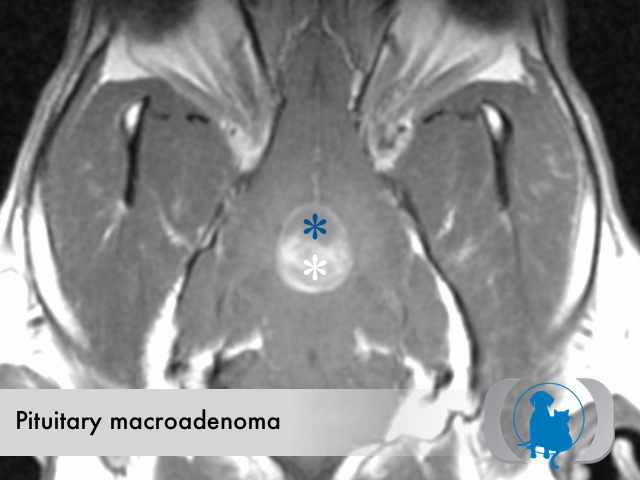
Dorsal T1 weighted post contrast magnetic resonance image (MRI) of the brain of a 6 year old neutered male Labrador Retriever dog with a partially cystic (blue asterisk) pituitary macroadenoma (white asterisk).
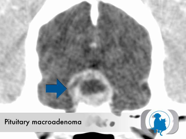
CT scan of a 6 year old neutered male Golden Retriever dog with a cystic pituitary macroadenoma.