Etiology
Ectopic ureters are a congenital condition that occur when the embryonic mesonephric and metanephric duct systems do not develop normally. The metanephric duct eventually develops into the ureter. In patients with ectopic ureters, the metanephric duct originates in an abnormal location or migrates in an abnormal fashion, resulting in malposition of the ureteral orifice either caudally within the urinary bladder or within the lumen of the urethra. Occasionally the ureter actually empties into the vagina or vestibule.
Pathophysiology
Urinary incontinence is the most common clinical manifestation of ectopic ureters. Incontinence occurs because the ureter enters the bladder distal to the normal location at the urinary bladder trigone leading to an ineffective urethral sphincter mechanism. Increased resistance to urine flow at the termination of the ureter can lead to increased ureteral pressure and secondary hydroureter and hydronephrosis which can lead to reduced renal function over time. Because the abnormal anatomy that leads to the symptoms is present at birth, the symptoms are generally recognized and the diagnosis achieved in young patients. Certain canine breeds have been found to be over represented namely:
Golden retriever
Labrador retriever
Miniature poodle
Newfoundland
Siberian husky
Soft-coated Wheaten terrier
Terriers
Toy poodle
West Highland white terrier
Clinical Signs
Urinary incontinence is the major symptom motivating presentation for evaluation.
Diagnostic Tests
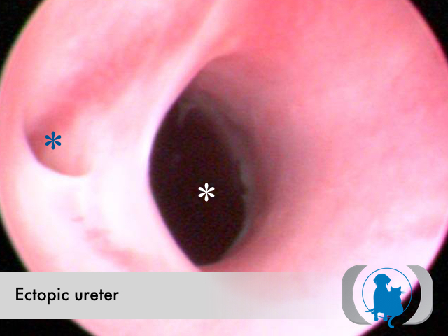
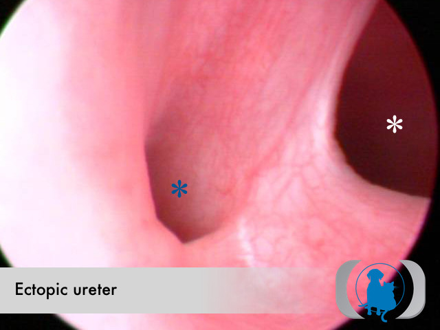
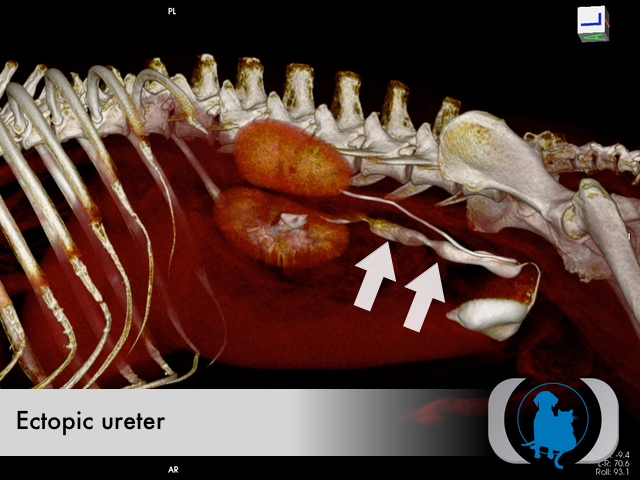
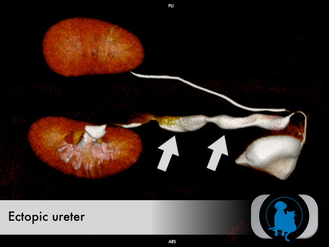
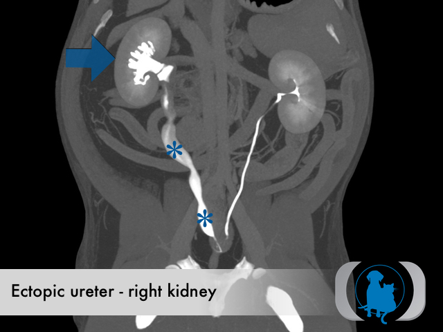
Urethroscopy/cystoscopy is the current gold standard test for diagnosing ectopic ureters, however contrast CT has recently been recognized for its ability to evaluate the entire renal system to identify both the abnormal local anatomy at the termination of the ureter as well as the effects on the entire length of the ureter and the kidney itself. Historically intravenous pyelography has been used but is limited by the 2 dimensional nature of the images obtained.
Therapy
Historically surgery has been the treatment of choice for ectopic ureters. More recently cystoscopy guided laser ablation has become possible for ectopic ureters with sufficient intramural tunneling.
Image Gallery