Etiology
The term hydrocephalus is derived from the greek "hydro" for water and "cephalus" for head. As such the condition is one in which there is an increased accumulation of cerebrospinal fluid (CSF) within the intracranial cavity. Hydrocephalus is considered congenital (sometimes called communicating) when the factors causing it are present at birth and acquired (sometimes called non-communicating) when it develops subsequent to conditions that develop thereafter.
Pathophysiology
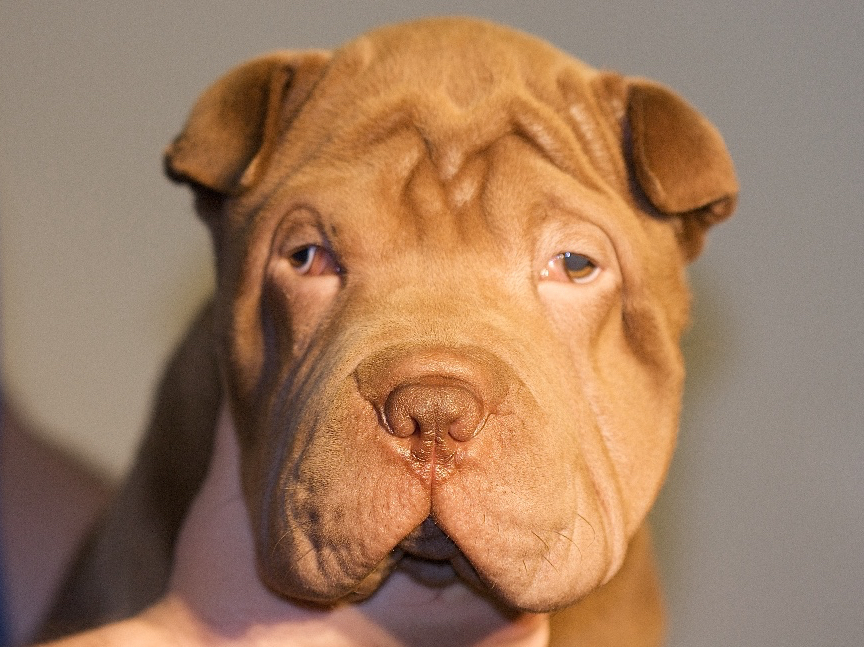
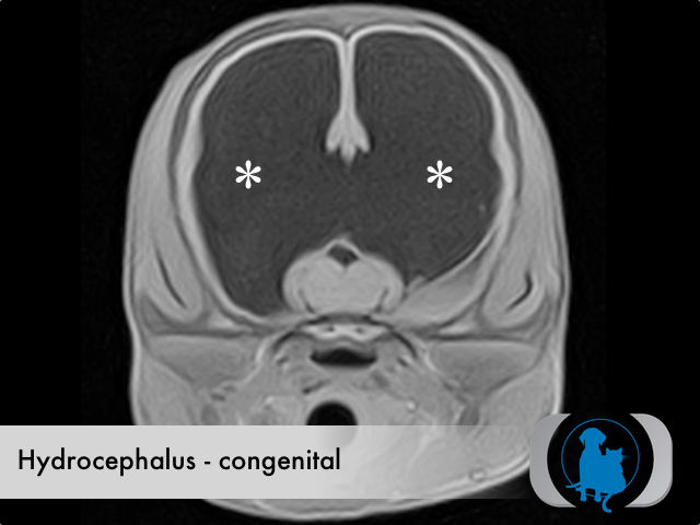
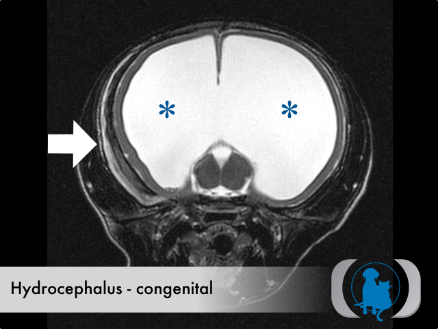
Hydrocephalus can result from either overproduction or reduced elimination of CSF from the intracranial cavity. Increased production of CSF is a form of acquired hydrocephalus and is uncommon, largely limited to tumors of the choroid plexus (the tissue within the ventricular system of the brain that is responsible for CSF production). Reduced elimination of CSF can be caused by tumors or infections that obstruct the path of CSF flow through and out of the intracranial cavity. These causes are readily identifiable by advanced imaging techniques like MRI and CT and represent a cause of acquired hydrocephalus. The underlying cause for congenital hydrocephalus is often harder to define but can be caused by macroscopic abnormalities (e.g., mesencephalic aqueduct stenosis) that obstruct CSF outflow as well as sub gross pathology that leads to reduced CSF absorption. The nature of this sub gross pathology is usually unknown. Congenital hydrocephalus is usually diagnosed in young patients. No sex predisposition is appreciated in either dogs or cats. While no breed disposition is noted in the cat. There are several canine breed predisposed including the Chihuahua, Yorkshire terrier, Manchester terrier, Toy Poodle, Boston terrier, Pekingese, and English bulldog.
Clinical Signs
The clinical signs of congenital hydrocephalus are usually secondary to the thinning of the surrounding cerebral cortex leading to progressive forebrain dysfunction. Common symptoms include dull mentation, head pressing, seizures, visual impairment, dementia and learning deficits. A common clinical finding of bilateral lateral strabismus is common in young dogs diagnosed with congenital hydrocephalus.
Diagnostic Tests
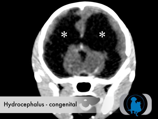
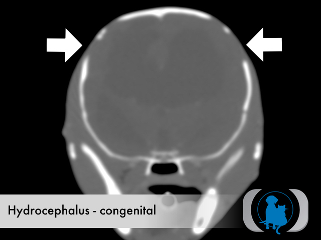
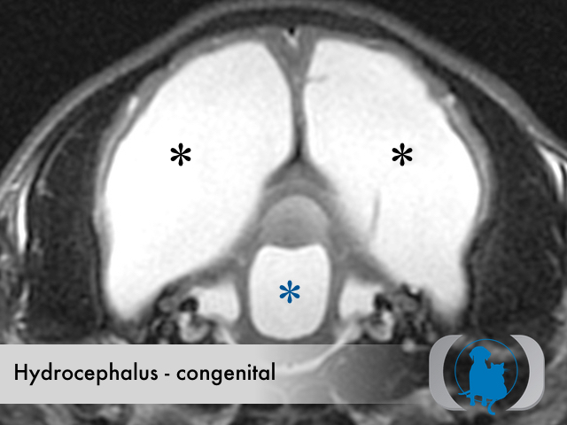
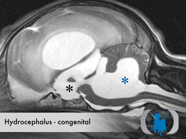
If diagnosed when fontanelles are open, ultrasound can be used to diagnose congenital hydrocephalus. When fontanelles are closed, advanced imaging with CT or MRI are needed to identify the enlarged ventricular system that develops.
Therapy
Medical management for congenital hydrocephalus includes the use of mannitol +/- furosemide to acutely lower intracranial pressure as well as prednisone, carbonic anhydrase inhibitors (e.g., acetazolamide, methazolamide) and potentially omeprazole to chronically reduce CSF production. Surgical therapy based on the placement of a ventriculoperitoneal shunt represents a more permanent solution.
Image Gallery